Which Organism is Unicellular and Possesses a Feeding Groove Group of Answer Choices
Excavata
Excavata, defined by a feeding groove that is "excavated" from one side, includes Diplomonads, Parabasalids and Euglenozoans.
Learning Objectives
Describe characteristics of Excavates, including Diplomonads, Parabasalids and Euglenozoans
Key Takeaways
Key Points
- Excavata are a supergroup of protists that are defined by an asymmetrical appearance with a feeding groove that is "excavated" from one side; it includes various types of organisms which are parasitic, photosynthetic and heterotrophic predators.
- Excavata includes the protists: Diplomonads, Parabasalids and Euglenozoans.
- Diplomonads are defined by the presence of a nonfunctional, mitochrondrial-remnant organelle called a mitosome.
- Parabasalids are characterized by a semi-functional mitochondria referred to as a hydrogenosome; they are comprised of parasitic protists, such as Trichomonas vaginalis.
- Euglenozoans can be classified as mixotrophs, heterotrophs, autotrophs, and parasites; they are defined by their use of flagella for movement.
Key Terms
- mitosome: an organelle found within certain unicellular eukaryotes which lack mitochondria
- hydrogenosome: a membrane-bound organelle found in ciliates, trichomonads, and fungi which produces molecular hydrogen and ATP
- kinetoplast: a disk-shaped mass of circular DNA inside a large mitochondrion, found specifically in protozoa of the class Kinetoplastea
Excavata
Many of the protist species classified into the supergroup Excavata are asymmetrical, single-celled organisms with a feeding groove "excavated" from one side. This supergroup includes heterotrophic predators, photosynthetic species, and parasites. Its subgroups are the diplomonads, parabasalids, and euglenozoans.
Diplomonads
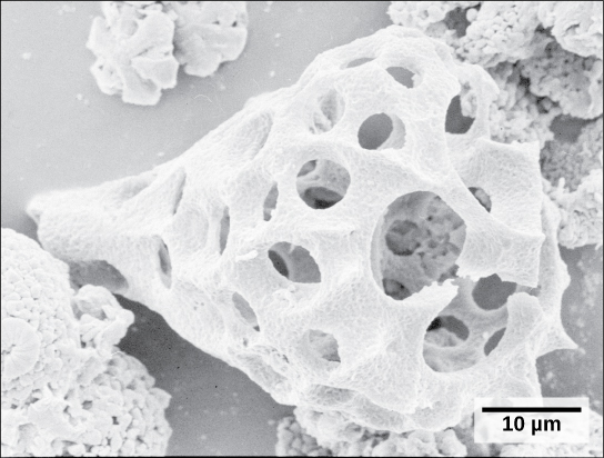
Among the Excavata are the diplomonads, which include the intestinal parasite, Giardia lamblia. Until recently, these protists were believed to lack mitochondria. Mitochondrial remnant organelles, called mitosomes, have since been identified in diplomonads, but these mitosomes are essentially nonfunctional. Diplomonads exist in anaerobic environments and use alternative pathways, such as glycolysis, to generate energy. Each diplomonad cell has two identical nuclei and uses several flagella for locomotion.
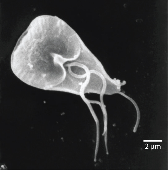
Giardia lamblia
The mammalian intestinal parasite Giardia lamblia, visualized here using scanning electron microscopy, is a waterborne protist that causes severe diarrhea when ingested.
Parabasalids
A second Excavata subgroup, the parabasalids, also exhibits semi-functional mitochondria. In parabasalids, these structures function anaerobically and are called hydrogenosomes because they produce hydrogen gas as a byproduct. Parabasalids move with flagella and membrane rippling. Trichomonas vaginalis, a parabasalid that causes a sexually-transmitted disease in humans, employs these mechanisms to transit through the male and female urogenital tracts. T. vaginalis causes trichomoniasis, which appears in an estimated 180 million cases worldwide each year. Whereas men rarely exhibit symptoms during an infection with this protist, infected women may become more susceptible to secondary infection with human immunodeficiency virus (HIV) or genital wart virus infection, which causes over 90% of cervical cancer. Pregnant women infected with T. vaginalis are at an increased risk of serious complications, such as pre-term delivery.
Euglenozoans
Euglenozoans includes parasites, heterotrophs, autotrophs, and mixotrophs, ranging in size from 10 to 500 µm. Euglenoids move through their aquatic habitats using two long flagella that guide them toward light sources sensed by a primitive ocular organ called an eyespot. The familiar genus, Euglena, encompasses some mixotrophic species that display a photosynthetic capability only when light is present. In the dark, the chloroplasts of Euglena shrink up and temporarily cease functioning; the cells, instead, take up organic nutrients from their environment.
The human parasite, Trypanosoma brucei, belongs to a different subgroup of Euglenozoa, the kinetoplastids. The kinetoplastid subgroup is named after the kinetoplast, a DNA mass carried within the single, oversized mitochondrion possessed by each of these cells. This subgroup includes several parasites, collectively called trypanosomes, which cause devastating human diseases by infecting an insect species during a portion of their life cycle. T. brucei develops in the gut of the tsetse fly after the fly bites an infected human or other mammalian host. The parasite then travels to the insect salivary glands to be transmitted to another human or other mammal when the infected tsetse fly consumes another blood meal. T. brucei is common in central Africa and is the causative agent of African sleeping sickness, a disease associated with severe chronic fatigue and coma; it can be fatal if left untreated.
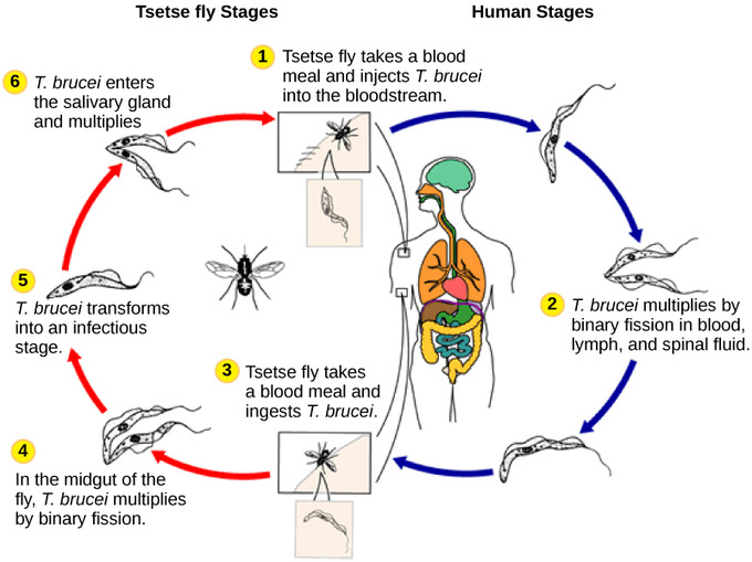
Life cycle of Trypanosoma brucei : Trypanosoma brucei, the causative agent of sleeping sickness, spends part of its life cycle in the tsetse fly and part in humans.
Chromalveolata: Alveolates
Alveolates are defined by the presence of an alveolus beneath the cell membrane and include dinoflagellates, apicomplexans and ciliates.
Learning Objectives
Evaluate traits associated with protists classified as alveolates which include dinoflagellates, apicomplexans, and ciliates
Key Takeaways
Key Points
- Alveolates are classified under the group Chromalveolata which developed as a result of a secondary endosymbiotic event.
- Dinoflagellates are defined by their flagella structure which lays perpendicular and fits into the cellulose plates of the dinoflagellate, promoting a spinning motion.
- Apicomplexans are defined by the asymmetrical distribution of their microtubules, fibrin, and vacuoles; they include the parasitic protist Plasmodium which causes malaria.
- Ciliates are defined by the presence of cilia (such as the oral groove in the Paramecium), which beat synchronously to aid the organism in locomotion and obtaining nutrients.
- Ciliates are defined by the presence of cilia, which beat synchronously, to aid the organism in locomotion and obtaining nutrients, such as the oral groove in the Paramecium.
Key Terms
- osmoregulation: the homeostatic regulation of osmotic pressure in the body in order to maintain a constant water content
- plastid: any of various organelles found in the cells of plants and algae, often concerned with photosynthesis
- conjugation: the temporary fusion of organisms, especially as part of sexual reproduction
Chromalveolata
Current evidence suggests that species classified as chromalveolates are derived from a common ancestor that engulfed a photosynthetic red algal cell, which itself had already evolved chloroplasts from an endosymbiotic relationship with a photosynthetic prokaryote. Therefore, the ancestor of chromalveolates is believed to have resulted from a secondary endosymbiotic event. However, some chromalveolates appear to have lost red alga-derived plastid organelles or lack plastid genes altogether. Therefore, this supergroup should be considered a hypothesis-based working group that is subject to change and can be subdivided into alveolates and stramenopiles.
Alveolates
A large body of data supports that the alveolates are derived from a shared common ancestor. The alveolates are named for the presence of an alveolus, or membrane-enclosed sac, beneath the cell membrane. The exact function of the alveolus is unknown, but it may be involved in osmoregulation. The alveolates are further categorized into the dinoflagellates, the apicomplexans, and the ciliates.
Dinoflagellates
Dinoflagellates exhibit extensive morphological diversity and can be photosynthetic, heterotrophic, or mixotrophic. Many dinoflagellates are encased in interlocking plates of cellulose with two perpendicular flagella that fit into the grooves between the cellulose plates. One flagellum extends longitudinally and a second encircles the dinoflagellate. Together, the flagella contribute to the characteristic spinning motion of dinoflagellates. These protists exist in freshwater and marine habitats; they are a component of plankton.
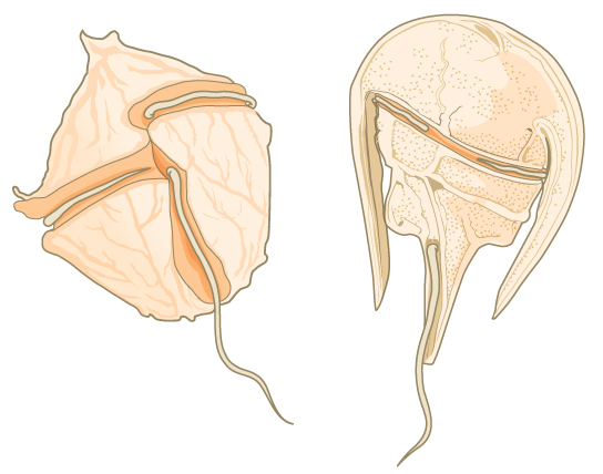
Dinoflagellates: The dinoflagellates exhibit great diversity in shape. Many are encased in cellulose armor and have two flagella that fit in grooves between the plates. Movement of these two perpendicular flagella causes a spinning motion.
Some dinoflagellates generate light, called bioluminescence, when they are jarred or stressed. Large numbers of marine dinoflagellates (billions or trillions of cells per wave) can emit light and cause an entire breaking wave to twinkle or take on a brilliant blue color. For approximately 20 species of marine dinoflagellates, population explosions (called blooms) during the summer months can tint the ocean with a muddy red color. This phenomenon is called a red tide and results from the abundant red pigments present in dinoflagellate plastids. In large quantities, these dinoflagellate species secrete an asphyxiating toxin that can kill fish, birds, and marine mammals. Red tides can be massively detrimental to commercial fisheries; humans who consume these protists may become poisoned.

Bioluminescence: Bioluminescence is emitted from dinoflagellates in a breaking wave, as seen from the New Jersey coast.
Apicomplexans
The apicomplexan protists are so named because their microtubules, fibrin, and vacuoles are asymmetrically distributed at one end of the cell in a structure called an apical complex. The apical complex is specialized for entry and infection of host cells. Indeed, all apicomplexans are parasitic. This group includes the genus Plasmodium, which causes malaria in humans. Apicomplexan life cycles are complex, involving multiple hosts and stages of sexual and asexual reproduction.
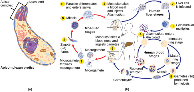
Parasitic apicomplexans: (a) Apicomplexans are parasitic protists. They have a characteristic apical complex that enables them to infect host cells. (b) Plasmodium, the causative agent of malaria, has a complex life cycle typical of apicomplexans.
Ciliates
The ciliates, which include Paramecium and Tetrahymena, are a group of protists 10 to 3,000 micrometers in length that are covered in rows, tufts, or spirals of tiny cilia. By beating their cilia synchronously or in waves, ciliates can coordinate directed movements and ingest food particles. Certain ciliates have fused cilia-based structures that function like paddles, funnels, or fins. Ciliates also are surrounded by a pellicle, providing protection without compromising agility. The genus Paramecium includes protists that have organized their cilia into a plate-like primitive mouth called an oral groove, which is used to capture and digest bacteria. Food captured in the oral groove enters a food vacuole where it combines with digestive enzymes. Waste particles are expelled by an exocytic vesicle that fuses at a specific region on the cell membrane: the anal pore. In addition to a vacuole-based digestive system, Paramecium also uses contractile vacuoles: osmoregulatory vesicles that fill with water as it enters the cell by osmosis and then contract to squeeze water from the cell.
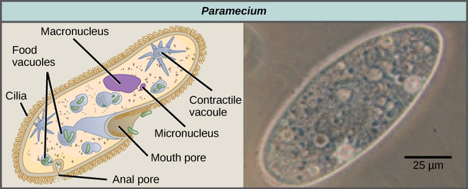
Paramecium: Paramecium has a primitive mouth (called an oral groove) to ingest food and an anal pore to excrete it. Contractile vacuoles allow the organism to excrete excess water. Cilia enable the organism to move.
Paramecium has two nuclei, a macronucleus and a micronucleus, in each cell. The micronucleus is essential for sexual reproduction, whereas the macronucleus directs asexual binary fission and all other biological functions. The process of sexual reproduction in Paramecium underscores the importance of the micronucleus to these protists. Paramecium and most other ciliates reproduce sexually by conjugation. This process begins when two different mating types of Paramecium make physical contact and join with a cytoplasmic bridge. The diploid micronucleus in each cell then undergoes meiosis to produce four haploid micronuclei. Three of these degenerate in each cell, leaving one micronucleus that then undergoes mitosis, generating two haploid micronuclei. The cells each exchange one of these haploid nuclei and move away from each other. A similar process occurs in bacteria that have plasmids. Fusion of the haploid micronuclei generates a completely novel diploid pre-micronucleus in each conjugative cell. This pre-micronucleus undergoes three rounds of mitosis to produce eight copies, while the original macronucleus disintegrates. Four of the eight pre-micronuclei become full-fledged micronuclei, whereas the other four perform multiple rounds of DNA replication and then become new macronuclei. Two cell divisions then yield four new paramecia from each original conjugative cell.
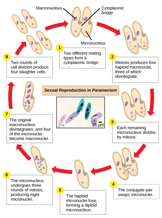
Paramecium: sexual reproduction: The complex process of sexual reproduction in Paramecium creates eight daughter cells from two original cells. Each cell has a macronucleus and a micronucleus. During sexual reproduction, the macronucleus dissolves and is replaced by a micronucleus.
Chromalveolata: Stramenopiles
Stramenophiles include photosynthetic marine algae and heterotrophic protists such as diatoms, brown and golden algae, and oomycetes.
Learning Objectives
Describe characteristics of the following Stramenophiles: diatoms, brown algae, golden algae, and oomycetes
Key Takeaways
Key Points
- Stramenophiles, also referred to as heterokonts, are a subclass of chromalveolata, and are identified by the presence of a "hairy" flagellum.
- Diatoms, present in both freshwater and marine plankton, are unicellular photosynthetic protists that are characterized by the presence of a cell wall composed of silicon dioxide that displays intricate patterns.
- Golden algae, present in both freshwater and marine plankton communities, are unicellular photosynthetic protists characterized by the presence of carotenoids (yellow-orange photosynthetic pigments).
- Oomycetes, commonly referred to as water molds, are characterized by their fungus-like morphology, a cellulose-based cell wall, and a filamentous network used for nutrient uptake.
- Oomycetes, commonly referred to as water molds, are characterized by their fungus-like morphology, a cellulose-based cell wall and a filamentous network used for nutrient uptake.
Key Terms
- stipe: the stem of a kelp
- raphe: a ridge or seam on an organ, bodily tissue, or other structure, especially at the join between two halves or sections
- saprobe: an organism that lives off of dead or decaying organic material
Chromalveolates
Current evidence suggests that chromalveolates have an ancestor which resulted from a secondary endosymbiotic event. The species which fall under the classification of chromalveolates have evolved from a common ancestor that engulfed a photosynthetic red algal cell. This red algal cell had previously evolved chloroplasts from an endosymbiotic relationship with a photosynthetic prokaryote. Chromalveolates include very important photosynthetic organisms, such as diatoms, brown algae, and significant disease agents in animals and plants. The chromalveolates can be subdivided into alveolates and stramenopiles.
Stramenopiles
A subgroup of chromalveolates, the stramenopiles, also referred to as heterokonts, includes photosynthetic marine algae and heterotrophic protists. The unifying feature of this group is the presence of a textured, or "hairy," flagellum. Many stramenopiles also have an additional flagellum that lacks hair-like projections. Members of this subgroup range in size from single-celled diatoms to the massive and multicellular kelp.
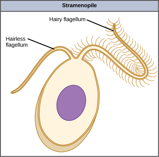
Stramenophile structure: This stramenopile cell has a single hairy flagellum and a secondary smooth flagellum.
Diatoms
The diatoms are unicellular photosynthetic protists that encase themselves in intricately patterned, glassy cell walls composed of silicon dioxide in a matrix of organic particles. These protists are a component of freshwater and marine plankton. Most species of diatoms reproduce asexually, although some instances of sexual reproduction and sporulation also exist. Some diatoms exhibit a slit in their silica shell called a raphe. By expelling a stream of mucopolysaccharides from the raphe, the diatom can attach to surfaces or propel itself in one direction.
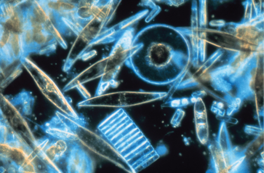
Diatoms: Assorted diatoms, visualized here using light microscopy, live among annual sea ice in McMurdo Sound, Antarctica. Diatoms range in size from 2 to 200 µm.
During periods of nutrient availability, diatom populations bloom to numbers greater than can be consumed by aquatic organisms. The excess diatoms die and sink to the sea floor where they are not easily reached by saprobes that feed on dead organisms. As a result, the carbon dioxide that the diatoms had consumed and incorporated into their cells during photosynthesis is not returned to the atmosphere. In general, this process by which carbon is transported deep into the ocean is described as the biological carbon pump because carbon is "pumped" to the ocean depths where it is inaccessible to the atmosphere as carbon dioxide. The biological carbon pump is a crucial component of the carbon cycle that helps to maintain lower atmospheric carbon dioxide levels.
Golden Algae
Like diatoms, golden algae are largely unicellular, although some species can form large colonies. Their characteristic gold color results from their extensive use of carotenoids, a group of photosynthetic pigments that are generally yellow or orange in color. Golden algae are found in both freshwater and marine environments, where they form a major part of the plankton community.
Brown Algae
The brown algae are primarily marine, multicellular organisms that are known colloquially as seaweeds. Giant kelps are a type of brown algae. Some brown algae have evolved specialized tissues that resemble terrestrial plants, with root-like holdfasts, stem-like stipes, and leaf-like blades that are capable of photosynthesis. The stipes of giant kelps are enormous, extending in some cases for 60 meters. A variety of algal life cycles exists, but the most complex is alternation of generations in which both haploid and diploid stages involve multicellularity. For instance, compare this life cycle to that of humans. In humans, haploid gametes produced by meiosis (sperm and egg) combine in fertilization to generate a diploid zygote that undergoes many rounds of mitosis to produce a multicellular embryo and then a fetus. However, the individual sperm and egg themselves never become multicellular beings. In the brown algae genus Laminaria, haploid spores develop into multicellular gametophytes, which produce haploid gametes that combine to produce diploid organisms that then become multicellular organisms with a different structure from the haploid form. Terrestrial plants also have evolved alternation of generations.
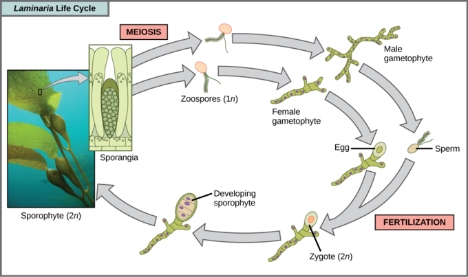
Brown algae life cycle: Several species of brown algae, such as the Laminaria shown here, have evolved life cycles in which both the haploid (gametophyte) and diploid (sporophyte) forms are multicellular. The gametophyte is different in structure from the sporophyte.
Oomycetes
The water molds, oomycetes ("egg fungus"), were so-named based on their fungus-like morphology, but molecular data have shown that the water molds are not closely related to fungi. The oomycetes are characterized by a cellulose-based cell wall and an extensive network of filaments that allow for nutrient uptake. As diploid spores, many oomycetes have two oppositely-directed flagella (one hairy and one smooth) for locomotion. The oomycetes are non-photosynthetic and include many saprobes and parasites. The saprobes appear as white fluffy growths on dead organisms. Most oomycetes are aquatic, but some parasitize terrestrial plants. One plant pathogen is Phytophthora infestans, the causative agent of late blight of potatoes, such as occurred in the nineteenth century Irish potato famine.
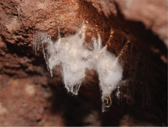
Oomycete: A saprobic oomycete engulfs a dead insect.
Rhizaria
Rhizaria are a supergroup of protists, typically amoebas, that are characterized by the presence of needle-like pseudopodia.
Learning Objectives
Describe characteristics associated with Rhizaria
Key Takeaways
Key Points
- The needle-like pseudopodia are used to carry out a process called cytoplasmic streaming which is a means of locomotion or distributing nutrients and oxygen.
- Two major subclassifications of Rhizaria include Forams and Radiolarians.
- Forams are characterized as unicellular heterotrophic protists that have porous shells, referred to as tests, which can contain photosynthetic algae that the foram can use as a nutrient source.
- Radiolarians are characterized by a glassy silica exterior that displays either bilateral or radial symmetry.
Key Terms
- pseudopodia: temporary projections of eukaryotic cells
- test: the external calciferous shell of a foram
Rhizaria
The Rhizaria supergroup includes many of the amoebas, most of which have threadlike or needle-like pseudopodia. Pseudopodia function to trap and engulf food particles and to direct movement in rhizarian protists. These pseudopods project outward from anywhere on the cell surface and can anchor to a substrate. The protist then transports its cytoplasm into the pseudopod, thereby moving the entire cell. This type of motion, called cytoplasmic streaming, is used by several diverse groups of protists as a means of locomotion or as a method to distribute nutrients and oxygen.
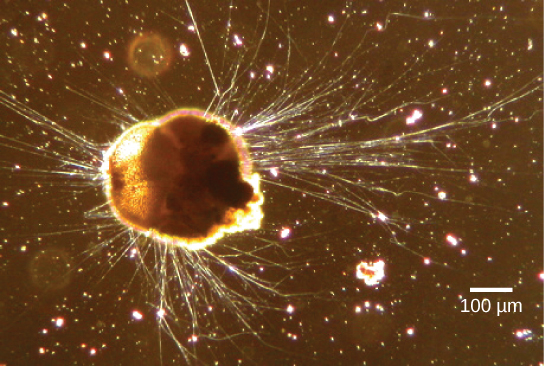
Ammonia tepida: Ammonia tepida, a Rhizaria species viewed here using phase contrast light microscopy, exhibits many threadlike pseudopodia.
Forams
Foraminiferans, or forams, are unicellular heterotrophic protists, ranging from approximately 20 micrometers to several centimeters in length; they occasionally resemble tiny snails. As a group, the forams exhibit porous shells, called tests, that are built from various organic materials and typically hardened with calcium carbonate. The tests may house photosynthetic algae, which the forams can harvest for nutrition. Foram pseudopodia extend through the pores and allow the forams to move, feed, and gather additional building materials. Foraminiferans are also useful as indicators of pollution and changes in global weather patterns.
The life-cycle involves an alternation between haploid and diploid phases. The haploid phase initially has a single nucleus, and divides to produce gametes with two flagella. The diploid phase is multinucleate, and after meiosis fragments to produce new organisms. The benthic forms has multiple rounds of asexual reproduction between sexual generations.
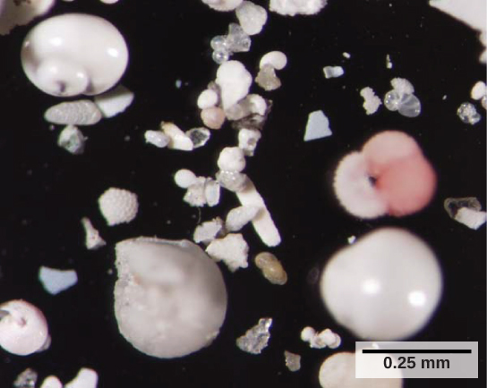
Forams: These shells from foraminifera sank to the sea floor.
Radiolarians
A second subtype of Rhizaria, the radiolarians, exhibit intricate exteriors of glassy silica with radial or bilateral symmetry. Radiolarians display needle-like pseudopods that are supported by microtubules which radiate outward from the cell bodies of these protists and function to catch food particles. The shells of dead radiolarians sink to the ocean floor, where they may accumulate in 100 meter-thick depths. Preserved, sedimented radiolarians are very common in the fossil record.
Radiolarian shell: This fossilized radiolarian shell was imaged using a scanning electron microscope.
Archaeplastida
Archaeplastida are a supergroup of protists that comprise red and green algae, which include unicellular, multicellular, and colonial forms.
Learning Objectives
Describe the relationship between red algae, green algae, and land plants
Key Takeaways
Key Points
- Archaeplastida are typically associated with their relationship to land plants; in addition, molecular evidence shows that Archaeplastida evolved from an endosymbiotic relationship between a heterotrophic protist and a cyanobacterium.
- Red algae (rhodophytes), are classified as Archaeplastida and are most often characterized by the presence of the red pigment phycoerythrin; however, there are red algae that lack phycoerythrins and can be classified as parasites.
- Red algae typically exist as multicellular protists that lack flagella; however, they can also exist as unicellular organisms.
- Green algae are the most abundant group of algae and can be further classified as chlorophytes and charophytes.
- Charophytes are the green algae which resemble land plants and are their closest living relative.
- Chlorophytes are the green algae which exhibit a wide range of forms; they can be unicellular, multicellular, or colonial.
Key Terms
- endosymbiotic: that lives within a body or cells of another organism
- plankton: a generic term for all the organisms that float in the sea
Archaeplastida
Red algae and green algae are included in the supergroup Archaeplastida. It is well documented that land plants evolved from a common ancestor of these protists; their closest relatives are found within this group. Molecular evidence supports that all Archaeplastida are descendants of an endosymbiotic relationship between a heterotrophic protist and a cyanobacterium. The red and green algae include unicellular, multicellular, and colonial forms
Red Algae
Red algae, or rhodophytes, are primarily multicellular, lack flagella, and range in size from microscopic, unicellular protists to large, multicellular forms grouped into the informal seaweed category. The red algae life cycle is an alternation of generations. Some species of red algae contain phycoerythrins, photosynthetic accessory pigments that are red in color and outcompete the green tint of chlorophyll, making these species appear as varying shades of red. Other protists classified as red algae lack phycoerythrins and are parasites. Red algae are common in tropical waters where they have been detected at depths of 260 meters. Other red algae exist in terrestrial or freshwater environments.
Green Algae: Chlorophytes and Charophytes
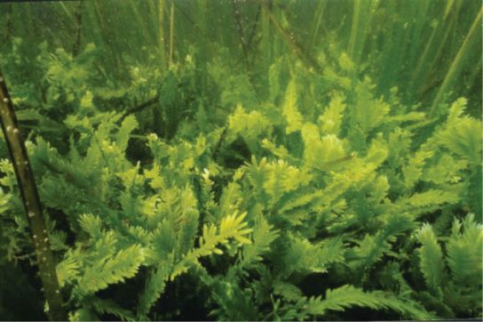
The most abundant group of algae is the green algae. The green algae exhibit similar features to the land plants, particularly in terms of chloroplast structure. It is well supported that this group of protists share a relatively-recent common ancestors with land plants. The green algae are subdivided into the chlorophytes and the charophytes. The charophytes are the closest-living relatives of land plants, resembling them in morphology and reproductive strategies. Charophytes are common in wet habitats where their presence often signals a healthy ecosystem.
The chlorophytes exhibit great diversity of form and function. Chlorophytes primarily inhabit freshwater and damp soil; they are a common component of plankton. Chlamydomonas is a simple, unicellular chlorophyte with a pear-shaped morphology and two opposing, anterior flagella that guide this protist toward light sensed by its eyespot. More complex chlorophyte species exhibit haploid gametes and spores that resemble Chlamydomonas.
The chlorophyte Volvox is one of only a few examples of a colonial organism, which behaves in some ways like a collection of individual cells, but in other ways like the specialized cells of a multicellular organism. Volvox colonies contain 500 to 60,000 cells, each with two flagella, contained within a hollow, spherical matrix composed of a gelatinous glycoprotein secretion. Individual Volvox cells move in a coordinated fashion and are interconnected by cytoplasmic bridges. Only a few of the cells reproduce to create daughter colonies, an example of basic cell specialization in this organism.
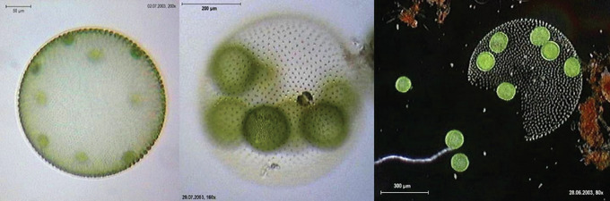
Volvox aureus
Volvox aureus is a green alga in the supergroup Archaeplastida. This species exists as a colony, consisting of cells immersed in a gel-like matrix and intertwined with each other via hair-like cytoplasmic extensions.
True multicellular organisms, such as the sea lettuce, Ulva, are represented among the chlorophytes. In addition, some chlorophytes exist as large, multinucleate, single cells. Species in the genus Caulerpa exhibit flattened, fern-like foliage and can reach lengths of 3 meters. Caulerpa species undergo nuclear division, but their cells do not complete cytokinesis, remaining instead as massive and elaborate single cells.
Caulerpa taxifolia
Caulerpa taxifolia is a chlorophyte consisting of a single cell containing potentially thousands of nuclei.
Amoebozoa and Opisthokonta
Amoebozoa are a type of protist that is characterized by the presence of pseudopodia which they use for locomotion and feeding.
Learning Objectives
Describe characteristics of Amoebozoa
Key Takeaways
Key Points
- Amoebozoa (amoebas) can live in either marine and fresh water or in soil.
- Amoebozoa are characterized by the presence of pseudopodia, which are extensions that can be either tube-like or flat lobes and are used for locomotion and feeding.
- Amooebozoa can be further divided into subclassifications that include slime molds; these can be found as both plasmodial and cellular types.
- Plasmodial slime molds are characterized by the presence of large, multinucleate cells that have the ability to glide along the surface and engulf food particles as they move.
- Cellular molds are characterized by the presence of independent amoeboid cells during times of nutrient abundancy and the development of a cellular mass, called a slug, during times of nutrient depletion.
- Archamoebae, Flabellinea, and Tubulinea are also groups of Amoebozoa; their defining characteristics include: Archamoebae lack mitochondria; Flabellinea flatten during locomotion and lack a shell and flagella; Tubulinea have a rough cylindrical form during locomotion with cylindrical pseudopodia.
Key Terms
- rhizaria: a species-rich supergroup of mostly unicellular eukaryotes that for the most part are amoeboids with filose, reticulose, or microtubule-supported pseudopods
- plasmodium: a mass of cytoplasm, containing many nuclei, created by the aggregation of amoeboid cells of slime molds during their vegetative phase
- sporangia: an enclosure in which spores are formed (also called a fruiting body)
Amoebozoa
Protists are eukaryotic organisms that are classified as unicellular, colonial, or multicellular organisms that do not have specialized tissues. This identifying property sets protists apart from other organisms within the Eukarya domain. The amoebozoans are classified as protists with pseudopodia which are used in locomotion and feeding. Amoebozoans live in marine environments, fresh water, or in soil. In addition to the defining pseudopodia, they also lack a shell and do not have a fixed body. The pseudopodia which are characteristically exhibited include extensions which can be tube-like or flat lobes, rather than the hair-like pseudopodia of rhizarian amoeba. Rhizarian amoeba are amoeboids with filose, reticulose, or microtubule-supported pseudopods and include the groups: Cercozoa, Foraminifera, and Radiolaria and are classified as bikonts. The Amoebozoa include several groups of unicellular amoeba-like organisms that are free-living or parasites that are classified as unikonts. The best known and most well-studied member of this group is the slime mold. Additional members include the Archamoebae, Tubulinea, and Flabellinea.
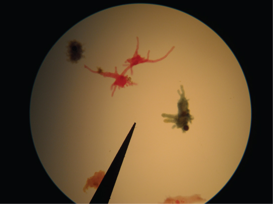
Pseudopodia structures: Amoebae with tubular and lobe-shaped pseudopodia, such as the ones seen under this microscope, would be morphologically classified as amoebozoans.
Slime Molds
A subset of the amoebozoans, the slime molds, has several morphological similarities to fungi that are thought to be the result of convergent evolution. For instance, during times of stress, some slime molds develop into spore -generating fruiting bodies, similar to fungi.
The slime molds are categorized on the basis of their life cycles into plasmodial or cellular types. Plasmodial slime molds are composed of large, multinucleate cells that move along surfaces like an amorphous blob of slime during their feeding stage. Food particles are lifted and engulfed into the slime mold as it glides along. Upon maturation, the plasmodium takes on a net-like appearance with the ability to form fruiting bodies, or sporangia, during times of stress. Haploid spores are produced by meiosis within the sporangia. These spores can be disseminated through the air or water to potentially land in more favorable environments. If this occurs, the spores germinate to form ameboid or flagellate haploid cells that can combine with each other and produce a diploid zygotic slime mold to complete the life cycle.

Badhamia utricularis: Badhamia utricularis: an example of a plasmodial slime mold with the ability to form a fruiting body.
The cellular slime molds function as independent amoeboid cells when nutrients are abundant. When food is depleted, cellular slime molds pile onto each other into a mass of cells that behaves as a single unit called a slug. Some cells in the slug contribute to a 2–3-millimeter stalk, drying up and dying in the process. Cells atop the stalk form an asexual fruiting body that contains haploid spores. As with plasmodial slime molds, the spores are disseminated and can germinate if they land in a moist environment. One representative genus of the cellular slime molds is Dictyostelium, which commonly exists in the damp soil of forests.

Plasmodial slime mold: Physarum polycephalum: Physarum polycephalum is an example of a cellular slime mold.
Archamoebae, Flabellinea, and Tubulinea
The Archamoebae are a group of Amoebozoa distinguished by the absence of mitochondria. They include genera that are internal parasites or commensals of animals (Entamoeba and Endolimax). A few species are human pathogens, causing diseases such as amoebic dysentery. The other genera of archamoebae live in freshwater habitats and are unusual among amoebae in possessing flagella. Most have a single nucleus and flagellum, but the giant amoeba, Pelomyxa, has many of each.
The Tubulinea are a major grouping of Amoebozoa, including most of the larger and more familiar amoebae like Amoeba, Arcella, and Difflugia. During locomotion, most Tubulinea have a roughly cylindrical form or produce numerous cylindrical pseudopods. Each cylinder advances by a single central stream of cytoplasm, granular in appearance, and has no subpseudopodia. This distinguishes them from other amoeboid groups, although in some members this is not the normal type of locomotion.
Licenses and Attributions
CC licensed content, Shared previously
- Curation and Revision. Provided by: Boundless.com. License: CC BY-SA: Attribution-ShareAlike
CC licensed content, Specific attribution
- OpenStax College, Biology. October 16, 2013. Provided by: OpenStax CNX. Located at: https://openstax.org/books/biology/pages/23-3-groups-of-protists. License: CC BY: Attribution
- kinetoplast. Provided by: Wiktionary. License: CC BY-SA: Attribution-ShareAlike
- hydrogenosome. Provided by: Wiktionary. License: CC BY-SA: Attribution-ShareAlike
- mitosome. Provided by: Wiktionary. License: CC BY-SA: Attribution-ShareAlike
- OpenStax College, Groups of Protists. October 16, 2013. Provided by: OpenStax CNX. Located at: https://cnx.org/resources/a28f470d0d864cfd1240440887eef49f3c415ea0/Figure_23_03_03.jpg. License: CC BY: Attribution
- OpenStax College, Groups of Protists. October 16, 2013. Provided by: OpenStax CNX. Located at: https://cnx.org/resources/2cb4261724af7afd5858a434724d0e14a3a11c21/Figure_23_03_02.jpg. License: CC BY: Attribution
- osmoregulation. Provided by: Wiktionary. License: CC BY-SA: Attribution-ShareAlike
- plastid. Provided by: Wiktionary. License: CC BY-SA: Attribution-ShareAlike
- OpenStax College, Biology. October 16, 2013. Provided by: OpenStax CNX. Located at: https://openstax.org/books/biology/pages/23-3-groups-of-protists. License: CC BY: Attribution
- conjugation. Provided by: Wiktionary. License: CC BY-SA: Attribution-ShareAlike
- OpenStax College, Groups of Protists. October 16, 2013. Provided by: OpenStax CNX. Located at: https://cnx.org/resources/a28f470d0d864cfd1240440887eef49f3c415ea0/Figure_23_03_03.jpg. License: CC BY: Attribution
- OpenStax College, Groups of Protists. October 16, 2013. Provided by: OpenStax CNX. Located at: https://cnx.org/resources/2cb4261724af7afd5858a434724d0e14a3a11c21/Figure_23_03_02.jpg. License: CC BY: Attribution
- OpenStax College, Groups of Protists. October 16, 2013. Provided by: OpenStax CNX. Located at: https://cnx.org/resources/34c735c435ea1311a6109ae66c1e8081474de7e7/Figure_23_03_04.jpg. License: CC BY: Attribution
- OpenStax College, Groups of Protists. October 16, 2013. Provided by: OpenStax CNX. Located at: https://cnx.org/resources/a8251696cbc1f006efbeccb9fa4461510c9e0440/Figure_23_03_05.jpg. License: CC BY: Attribution
- OpenStax College, Groups of Protists. October 16, 2013. Provided by: OpenStax CNX. Located at: https://cnx.org/resources/42809f1f90c60d1188604557f643841b4117be78/Figure_23_03_07.png. License: CC BY: Attribution
- OpenStax College, Groups of Protists. October 16, 2013. Provided by: OpenStax CNX. License: CC BY: Attribution
- OpenStax College, Groups of Protists. October 16, 2013. Provided by: OpenStax CNX. License: CC BY: Attribution
- OpenStax College, Biology. October 16, 2013. Provided by: OpenStax CNX. Located at: https://openstax.org/books/biology/pages/23-3-groups-of-protists. License: CC BY: Attribution
- raphe. Provided by: Wiktionary. License: CC BY-SA: Attribution-ShareAlike
- saprobe. Provided by: Wiktionary. License: CC BY-SA: Attribution-ShareAlike
- stipe. Provided by: Wiktionary. License: CC BY-SA: Attribution-ShareAlike
- OpenStax College, Groups of Protists. October 16, 2013. Provided by: OpenStax CNX. Located at: https://cnx.org/resources/a28f470d0d864cfd1240440887eef49f3c415ea0/Figure_23_03_03.jpg. License: CC BY: Attribution
- OpenStax College, Groups of Protists. October 16, 2013. Provided by: OpenStax CNX. Located at: https://cnx.org/resources/2cb4261724af7afd5858a434724d0e14a3a11c21/Figure_23_03_02.jpg. License: CC BY: Attribution
- OpenStax College, Groups of Protists. October 16, 2013. Provided by: OpenStax CNX. Located at: https://cnx.org/resources/34c735c435ea1311a6109ae66c1e8081474de7e7/Figure_23_03_04.jpg. License: CC BY: Attribution
- OpenStax College, Groups of Protists. October 16, 2013. Provided by: OpenStax CNX. Located at: https://cnx.org/resources/a8251696cbc1f006efbeccb9fa4461510c9e0440/Figure_23_03_05.jpg. License: CC BY: Attribution
- OpenStax College, Groups of Protists. October 16, 2013. Provided by: OpenStax CNX. Located at: https://cnx.org/resources/42809f1f90c60d1188604557f643841b4117be78/Figure_23_03_07.png. License: CC BY: Attribution
- OpenStax College, Groups of Protists. October 16, 2013. Provided by: OpenStax CNX. License: CC BY: Attribution
- OpenStax College, Groups of Protists. October 16, 2013. Provided by: OpenStax CNX. License: CC BY: Attribution
- OpenStax College, Groups of Protists. October 16, 2013. Provided by: OpenStax CNX. Located at: https://cnx.org/resources/084a16cfbc6388a3acef8f802f0bc843b3195237/Figure_23_03_09.jpg. License: CC BY: Attribution
- OpenStax College, Groups of Protists. October 16, 2013. Provided by: OpenStax CNX. Located at: https://cnx.org/resources/752662c68c1ba7b4ebf94ef63e3c260bc8373a4b/Figure_23_03_10.png. License: CC BY: Attribution
- OpenStax College, Groups of Protists. October 16, 2013. Provided by: OpenStax CNX. Located at: https://cnx.org/resources/4e5a900366bb8acdf3f7ff5fcda1c8a5b4d8b33d/Figure_23_03_08.jpg. License: CC BY: Attribution
- OpenStax College, Groups of Protists. October 16, 2013. Provided by: OpenStax CNX. Located at: https://cnx.org/resources/d530fa26998f6135a932413fb6b167fb9c0683c9/Figure_23_03_11.jpg. License: CC BY: Attribution
- OpenStax College, Biology. October 16, 2013. Provided by: OpenStax CNX. Located at: https://openstax.org/books/biology/pages/23-3-groups-of-protists. License: CC BY: Attribution
- test. Provided by: Wiktionary. License: CC BY-SA: Attribution-ShareAlike
- pseudopodia. Provided by: Wikipedia. License: CC BY-SA: Attribution-ShareAlike
- OpenStax College, Groups of Protists. October 16, 2013. Provided by: OpenStax CNX. Located at: https://cnx.org/resources/a28f470d0d864cfd1240440887eef49f3c415ea0/Figure_23_03_03.jpg. License: CC BY: Attribution
- OpenStax College, Groups of Protists. October 16, 2013. Provided by: OpenStax CNX. Located at: https://cnx.org/resources/2cb4261724af7afd5858a434724d0e14a3a11c21/Figure_23_03_02.jpg. License: CC BY: Attribution
- OpenStax College, Groups of Protists. October 16, 2013. Provided by: OpenStax CNX. Located at: https://cnx.org/resources/34c735c435ea1311a6109ae66c1e8081474de7e7/Figure_23_03_04.jpg. License: CC BY: Attribution
- OpenStax College, Groups of Protists. October 16, 2013. Provided by: OpenStax CNX. Located at: https://cnx.org/resources/a8251696cbc1f006efbeccb9fa4461510c9e0440/Figure_23_03_05.jpg. License: CC BY: Attribution
- OpenStax College, Groups of Protists. October 16, 2013. Provided by: OpenStax CNX. Located at: https://cnx.org/resources/42809f1f90c60d1188604557f643841b4117be78/Figure_23_03_07.png. License: CC BY: Attribution
- OpenStax College, Groups of Protists. October 16, 2013. Provided by: OpenStax CNX. License: CC BY: Attribution
- OpenStax College, Groups of Protists. October 16, 2013. Provided by: OpenStax CNX. License: CC BY: Attribution
- OpenStax College, Groups of Protists. October 16, 2013. Provided by: OpenStax CNX. Located at: https://cnx.org/resources/084a16cfbc6388a3acef8f802f0bc843b3195237/Figure_23_03_09.jpg. License: CC BY: Attribution
- OpenStax College, Groups of Protists. October 16, 2013. Provided by: OpenStax CNX. Located at: https://cnx.org/resources/752662c68c1ba7b4ebf94ef63e3c260bc8373a4b/Figure_23_03_10.png. License: CC BY: Attribution
- OpenStax College, Groups of Protists. October 16, 2013. Provided by: OpenStax CNX. Located at: https://cnx.org/resources/4e5a900366bb8acdf3f7ff5fcda1c8a5b4d8b33d/Figure_23_03_08.jpg. License: CC BY: Attribution
- OpenStax College, Groups of Protists. October 16, 2013. Provided by: OpenStax CNX. Located at: https://cnx.org/resources/d530fa26998f6135a932413fb6b167fb9c0683c9/Figure_23_03_11.jpg. License: CC BY: Attribution
- OpenStax College, Groups of Protists. October 16, 2013. Provided by: OpenStax CNX. Located at: https://cnx.org/resources/a0eeea31926a4ead1b89589ccadc765eff845ff0/Figure_23_03_12.jpg. License: CC BY: Attribution
- OpenStax College, Groups of Protists. October 16, 2013. Provided by: OpenStax CNX. Located at: https://cnx.org/resources/5007e04b6177797f04d4ade363d111c1262bbab9/Figure_23_03_13.jpg. License: CC BY: Attribution
- OpenStax College, Groups of Protists. October 16, 2013. Provided by: OpenStax CNX. Located at: https://cnx.org/resources/b531f7738ffe977716711eedb37005c1fe42626a/Figure_23_03_14.jpg. License: CC BY: Attribution
- OpenStax College, Biology. October 16, 2013. Provided by: OpenStax CNX. Located at: https://openstax.org/books/biology/pages/23-3-groups-of-protists. License: CC BY: Attribution
- endosymbiotic. Provided by: Wiktionary. License: CC BY-SA: Attribution-ShareAlike
- plankton. Provided by: Wiktionary. License: CC BY-SA: Attribution-ShareAlike
- OpenStax College, Groups of Protists. October 16, 2013. Provided by: OpenStax CNX. Located at: https://cnx.org/resources/a28f470d0d864cfd1240440887eef49f3c415ea0/Figure_23_03_03.jpg. License: CC BY: Attribution
- OpenStax College, Groups of Protists. October 16, 2013. Provided by: OpenStax CNX. Located at: https://cnx.org/resources/2cb4261724af7afd5858a434724d0e14a3a11c21/Figure_23_03_02.jpg. License: CC BY: Attribution
- OpenStax College, Groups of Protists. October 16, 2013. Provided by: OpenStax CNX. Located at: https://cnx.org/resources/34c735c435ea1311a6109ae66c1e8081474de7e7/Figure_23_03_04.jpg. License: CC BY: Attribution
- OpenStax College, Groups of Protists. October 16, 2013. Provided by: OpenStax CNX. Located at: https://cnx.org/resources/a8251696cbc1f006efbeccb9fa4461510c9e0440/Figure_23_03_05.jpg. License: CC BY: Attribution
- OpenStax College, Groups of Protists. October 16, 2013. Provided by: OpenStax CNX. Located at: https://cnx.org/resources/42809f1f90c60d1188604557f643841b4117be78/Figure_23_03_07.png. License: CC BY: Attribution
- OpenStax College, Groups of Protists. October 16, 2013. Provided by: OpenStax CNX. License: CC BY: Attribution
- OpenStax College, Groups of Protists. October 16, 2013. Provided by: OpenStax CNX. License: CC BY: Attribution
- OpenStax College, Groups of Protists. October 16, 2013. Provided by: OpenStax CNX. Located at: https://cnx.org/resources/084a16cfbc6388a3acef8f802f0bc843b3195237/Figure_23_03_09.jpg. License: CC BY: Attribution
- OpenStax College, Groups of Protists. October 16, 2013. Provided by: OpenStax CNX. Located at: https://cnx.org/resources/752662c68c1ba7b4ebf94ef63e3c260bc8373a4b/Figure_23_03_10.png. License: CC BY: Attribution
- OpenStax College, Groups of Protists. October 16, 2013. Provided by: OpenStax CNX. Located at: https://cnx.org/resources/4e5a900366bb8acdf3f7ff5fcda1c8a5b4d8b33d/Figure_23_03_08.jpg. License: CC BY: Attribution
- OpenStax College, Groups of Protists. October 16, 2013. Provided by: OpenStax CNX. Located at: https://cnx.org/resources/d530fa26998f6135a932413fb6b167fb9c0683c9/Figure_23_03_11.jpg. License: CC BY: Attribution
- OpenStax College, Groups of Protists. October 16, 2013. Provided by: OpenStax CNX. Located at: https://cnx.org/resources/a0eeea31926a4ead1b89589ccadc765eff845ff0/Figure_23_03_12.jpg. License: CC BY: Attribution
- OpenStax College, Groups of Protists. October 16, 2013. Provided by: OpenStax CNX. Located at: https://cnx.org/resources/5007e04b6177797f04d4ade363d111c1262bbab9/Figure_23_03_13.jpg. License: CC BY: Attribution
- OpenStax College, Groups of Protists. October 16, 2013. Provided by: OpenStax CNX. Located at: https://cnx.org/resources/b531f7738ffe977716711eedb37005c1fe42626a/Figure_23_03_14.jpg. License: CC BY: Attribution
- OpenStax College, Groups of Protists. October 16, 2013. Provided by: OpenStax CNX. Located at: https://cnx.org/resources/1c3376c063d12309f6cfbaf4fd829cb8281dc212/Figure_23_03_16.jpg. License: CC BY: Attribution
- OpenStax College, Groups of Protists. October 16, 2013. Provided by: OpenStax CNX. Located at: https://cnx.org/resources/db00cc4b98cc4e392144aaf46138628225b82dc1/Figure_23_03_15.jpg. License: CC BY: Attribution
- OpenStax College, Biology. October 16, 2013. Provided by: OpenStax CNX. Located at: https://openstax.org/books/biology/pages/23-3-groups-of-protists. License: CC BY: Attribution
- Structural Biochemistry/Genome Analysis/Sequenced Genomes. Provided by: Wikibooks. License: CC BY-SA: Attribution-ShareAlike
- plasmodium. Provided by: Wiktionary. License: CC BY-SA: Attribution-ShareAlike
- sporangia. Provided by: Wikipedia. License: CC BY-SA: Attribution-ShareAlike
- Tubulinea. Provided by: Wikipedia. License: CC BY-SA: Attribution-ShareAlike
- OpenStax College, Biology. October 23, 2013. Provided by: OpenStax CNX. Located at: https://openstax.org/books/biology/pages/23-3-groups-of-protists. License: CC BY: Attribution
- Structural Biochemistry/Three Domains of Life/Eukarya. Provided by: Wikibooks. License: CC BY-SA: Attribution-ShareAlike
- Flabellinea. Provided by: Wikipedia. License: CC BY-SA: Attribution-ShareAlike
- Archamoebae. Provided by: Wikipedia. License: CC BY-SA: Attribution-ShareAlike
- Rhizaria. Provided by: Wikipedia. License: CC BY-SA: Attribution-ShareAlike
- rhizaria. Provided by: Wikipedia. License: CC BY-SA: Attribution-ShareAlike
- OpenStax College, Groups of Protists. October 16, 2013. Provided by: OpenStax CNX. Located at: https://cnx.org/resources/a28f470d0d864cfd1240440887eef49f3c415ea0/Figure_23_03_03.jpg. License: CC BY: Attribution
- OpenStax College, Groups of Protists. October 16, 2013. Provided by: OpenStax CNX. Located at: https://cnx.org/resources/2cb4261724af7afd5858a434724d0e14a3a11c21/Figure_23_03_02.jpg. License: CC BY: Attribution
- OpenStax College, Groups of Protists. October 16, 2013. Provided by: OpenStax CNX. Located at: https://cnx.org/resources/34c735c435ea1311a6109ae66c1e8081474de7e7/Figure_23_03_04.jpg. License: CC BY: Attribution
- OpenStax College, Groups of Protists. October 16, 2013. Provided by: OpenStax CNX. Located at: https://cnx.org/resources/a8251696cbc1f006efbeccb9fa4461510c9e0440/Figure_23_03_05.jpg. License: CC BY: Attribution
- OpenStax College, Groups of Protists. October 16, 2013. Provided by: OpenStax CNX. Located at: https://cnx.org/resources/42809f1f90c60d1188604557f643841b4117be78/Figure_23_03_07.png. License: CC BY: Attribution
- OpenStax College, Groups of Protists. October 16, 2013. Provided by: OpenStax CNX. License: CC BY: Attribution
- OpenStax College, Groups of Protists. October 16, 2013. Provided by: OpenStax CNX. License: CC BY: Attribution
- OpenStax College, Groups of Protists. October 16, 2013. Provided by: OpenStax CNX. Located at: https://cnx.org/resources/084a16cfbc6388a3acef8f802f0bc843b3195237/Figure_23_03_09.jpg. License: CC BY: Attribution
- OpenStax College, Groups of Protists. October 16, 2013. Provided by: OpenStax CNX. Located at: https://cnx.org/resources/752662c68c1ba7b4ebf94ef63e3c260bc8373a4b/Figure_23_03_10.png. License: CC BY: Attribution
- OpenStax College, Groups of Protists. October 16, 2013. Provided by: OpenStax CNX. Located at: https://cnx.org/resources/4e5a900366bb8acdf3f7ff5fcda1c8a5b4d8b33d/Figure_23_03_08.jpg. License: CC BY: Attribution
- OpenStax College, Groups of Protists. October 16, 2013. Provided by: OpenStax CNX. Located at: https://cnx.org/resources/d530fa26998f6135a932413fb6b167fb9c0683c9/Figure_23_03_11.jpg. License: CC BY: Attribution
- OpenStax College, Groups of Protists. October 16, 2013. Provided by: OpenStax CNX. Located at: https://cnx.org/resources/a0eeea31926a4ead1b89589ccadc765eff845ff0/Figure_23_03_12.jpg. License: CC BY: Attribution
- OpenStax College, Groups of Protists. October 16, 2013. Provided by: OpenStax CNX. Located at: https://cnx.org/resources/5007e04b6177797f04d4ade363d111c1262bbab9/Figure_23_03_13.jpg. License: CC BY: Attribution
- OpenStax College, Groups of Protists. October 16, 2013. Provided by: OpenStax CNX. Located at: https://cnx.org/resources/b531f7738ffe977716711eedb37005c1fe42626a/Figure_23_03_14.jpg. License: CC BY: Attribution
- OpenStax College, Groups of Protists. October 16, 2013. Provided by: OpenStax CNX. Located at: https://cnx.org/resources/1c3376c063d12309f6cfbaf4fd829cb8281dc212/Figure_23_03_16.jpg. License: CC BY: Attribution
- OpenStax College, Groups of Protists. October 16, 2013. Provided by: OpenStax CNX. Located at: https://cnx.org/resources/db00cc4b98cc4e392144aaf46138628225b82dc1/Figure_23_03_15.jpg. License: CC BY: Attribution
- OpenStax College, Groups of Protists. October 16, 2013. Provided by: OpenStax CNX. Located at: https://cnx.org/resources/7898d04061994cddadde19a4f3964a17ec29b917/Figure_23_03_17.jpg. License: CC BY: Attribution
- Fuligo92-300. Provided by: Wikipedia. License: CC BY: Attribution
- Badhamia utricularis mature. Provided by: Wikipedia. License: CC BY: Attribution
curreyhatterouble49.blogspot.com
Source: https://www.coursehero.com/study-guides/boundless-biology/groups-of-protists/